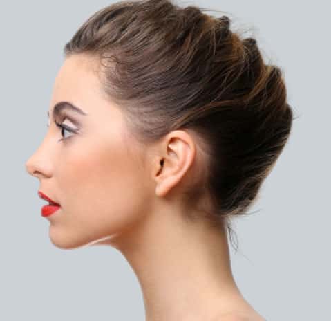
Ear Reconstruction Surgery in Mumbai
The hearing ability of humans is made possible by the presence of ears, which also play a significant part in facial attractiveness. External ear deformities and abnormalities are instantly noticeable and can also impair hearing. Ear reconstruction surgery in Mumbai, performed by Dr. Parag Telang at Designer Bodyz, can help regain natural-looking ears and lost confidence.
One of the most ear deformity people face is microtia, otoplasty, or losing their normal ear after trauma, burn, or injury. Microtia and lost ear can be reconstructed with microtia ear reconstruction surgery performed by Dr. Parag Telang, microtia ear surgeon in Mumbai at Designer Bodyz.
What is Microtia?
Microtia, or "little ear," refers to noticeable external ear abnormalities or deformities. For example, a congenital disorder in which the ear does not develop normally, resulting in small, oddly shaped, or occasionally absent ears, is called microtia. These abnormalities might impact both the internal and entire external ears. Sound, music, and speech cannot get to the inner ear and brain because of a narrower ear canal or an utterly closed ear canal, which can impair hearing. Ear reconstruction surgery in Mumbai is performed by Dr. Parag Telang can help to correct this issue and get back normal looking ears.
What Causes Microtia?
- Microtia often appears in the first few weeks of development during the first trimester of pregnancy. Although there is no established explanation for microtia, it has related to environmental factors, a diet low in carbohydrates and folic acid, genetic changes or disorders, and alcohol or drug use during pregnancy.
- Pregnancy-related use of acne medications is a known risk factor for microtia.
- A decrease in oxygen levels during the first trimester prevents the ear from developing, and multiple congenital disabilities are connected to acne medications used during pregnancy.
- Another potential risk factor for microtia in children is prenatal diabetes in the mother. Compared to other non-diabetic women, a woman with diabetes appears to have an increased probability of giving birth to a child with microtia.
- Even though microtia is not typically inherited, it can be in rare instances, and the disorder can skip generations.

Grades of Microtia
- Grade 1: Small ears and an ear canal that is frequently constricted or occasionally absent are characteristics of Grade 1.
- Grade 2: The upper half of the outer ear, which is often abnormally formed in Grade 2, frequently has a constricted or occasionally absent ear canal.
- Grade 3: The most prevalent ear ailment, Grade 3, is characterized by small, improperly formed ears without an ear canal.
- Grade 4: The missing ear and ear canal condition are Grade 4; anotia is another name for Grade 4 ear deformity.
Microtia's Impact on Life
Many children frequently experience diminished hearing because of a constricted or absent ear canal. When a person can only hear with one ear, the brain often struggles to block out background noise and cannot identify the sounds' origin and direction. Speech impairment in microtia patients generally results from diminished hearing in social and educational contexts.
The Perfect Age for Ear Reconstruction Surgery
Since the opposite (normal) ear stops growing in size after this age, nine years old and up is the optimal age for performing surgery to repair a nonexistent external ear. Therefore, the rib cartilage must be large enough for ear reconstruction surgery to build a complete ear framework.
Stages of Ear Reconstruction Surgery
Two stages are involved in ear reconstruction surgery in Mumbai that is performed at Designer Bodyz by the expert ear reconstruction doctor in India, Dr. Parag Telang.
Rib cartilage is removed in the initial stage. Harvested cartilage is used to build a three-dimensional scaffold for the ear. A replacement ear is made of cartilage using the second healthy ear of the patient as a template. Then the full ear framework is built and placed in a proper location on the side of the head.
From the side, the ear appears entire, although the sulcus is absent. Between three and six months following the first stage, the second stage is when the sulcus behind the ear is formed. For 5-7 days, a tight bandage keeps the skin graft in place.
The purpose of ear reconstruction surgery using rib cartilage is to give the underdeveloped ear the best possible structure and functionality. Reconstructive surgery on the ear canal cannot be performed on an infant. However, some families decide against surgical intervention as well. Surgery for ear reconstruction is frequently simpler in older kids since there is more cartilage available for grafting.
Rib cartilage graft surgery is advised for children between the ages of 8 and 10. Rib cartilage from the child's chest is taken out during rib cartilage graft surgery and used to restore the shape of an ear. Then the device is inserted beneath the skin where the ear would have been. After the new cartilage has fully absorbed at that location, more procedures and skin grafts may be necessary to place the ear better. Additionally, the implanted rib cartilage will feel more rigid and hard than the ear cartilage.
Advantages of Rib Cartilage Ear Reconstruction Surgery:
For people with microtia, rib cartilage ear restoration has the following benefits
- The procedure makes sole use of bodily tissue
- It is a tried-and-true method for ear reconstruction
- The patient may experience a fantastic, long-lasting outcome under skilled hands
- Rib cartilage ears can survive the rigors of most sports after surgery. Protective helmets are advised for contact sports like wrestling or football.
Disadvantages of Rib Cartilage Ear Construction Surgery:
For those with microtia, the drawbacks of rib cartilage repair include the following
- Removing rib cartilage hurts, necessitates one or two hospital nights, requires intravenous pain relievers, and leaves a visible chest scar
- A deformed chest wall may result from the removal of rib cartilage. Hence an expert surgeon must perform this surgery
- Hearing restoration is frequently postponed for years because atresia (ear canal) surgery cannot start until the ear reconstruction is complete
- Surgery may be required to "pin back" the average ear to make it more resemble the reconstructed ear
- Due to the cartilage frame's placement beneath the scalp, an ear that is "hairy" has a low scalp hairline.
Another popular ear reconstruction surgery performed to correct protruding ears in Mumbai is Otoplasty.
Otoplasty for Correcting Prominent Ears
A procedure to change the ear's shape, size, or position is termed "Otoplasty," a cosmetic ear surgery performed in Mumbai by Dr. Parag Telang. After the ears have gotten their entire length, otoplasty can be done at any age. Otoplasty can be done on both ears to optimize symmetry. Unfortunately, otoplasty won't alter the ability to hear or change the location of the ears.
Prominent Ears
There's no shortage of skin or cartilage in the prominent ear deformity. It is perfectly amenable to correction with ear molds. No matter their size, external ears (pinnae) protrude from the head's sides and are referred to as prominent ears. A prominent ear defines external ears (pinnae), which protrude from the head's sides, regardless of their size. The size of the external ear is less than 2 cm long and forms an angle of fewer than 25 degrees with respect to the head side; this abnormal appearance exceeds the regular head-to-ear measurements. The most common causes are defect, deformity, and abnormality in the occurrence of prominent ears, which can occur individually or in combination:
- Prominent concha deformity is caused either by an extensive concha-mastoid angle or an excessively deep concha. These two abnormalities can occur in combination and produce the most significant, deepest concavity of the pinna (prominent concha), which makes the center portion of the external ear prominent.
- Under-developed anti-helical fold: This deformity causes the protrusion of the helical rim and scapha, which occurs due to the inadequate folding of the anti-helix. This defect is manifested by the prominence of the elongated depression, which separates the helix, the anti-helix (scapha), the upper third of the upper ear, and occasionally the middle third ear.
- Protruding earlobe: Prominence of the lower third of the pinna is caused by this type of deformity of the earlobe.
Why is Otoplasty Done?
- If an ear or ears stick out too far from the head.
- If a person is dissatisfied with previous ear surgery.
- If the ears are large in proportion to the head.
If one is looking for otoplasty surgery in Mumbai, consult now Dr. Parag Telang at Designer Bodyz.
Otoplasty Procedure
It is used to set back ears so that they appear naturally proportionate and contoured without indication that surgical correction is the corrective goal of the otoplasty.
Procedure
- Based on what kind of correction is needed, the otoplasty procedure varies
- The doctor makes incisions on the back of the ear and within the inner creases of the ears
- A doctor might remove the excess skin and cartilage
- After that, fold the cartilage into the appropriate position and secure it from the inside
- The stitches will be closed with further stitches
- The whole process takes between two and three hours
- Following otoplasty, bandages will be placed around the ears for support and protection.
Otoplasty Pre-Operative Care
- The surgeon examines the condition of the ear and discusses all the related concerns
- For documentation purposes, the surgeon photographs the ear
- The patient must avoid vitamins, blood thinners, and anti-inflammatory medicines because they increase the risk of bleeding during surgery
- The patient should avoid smoking and drinking for a few weeks before and after surgery
- Consuming alcohol, cigarettes, and smoking can all delay blood flow and healing.
Otoplasty Post-Operative Care
- If the person feels discomfort or itching, they should take medications as the doctor recommend
- To prevent pressure on the ears, avoid sleeping on the surgical site
- Bandage will be removed after a few days of otoplasty. Ears are prone to swell and get red
- Cover the ears at night with a loose headband for at least two to six weeks
- Please speak with the doctor about when or whether sutures will be removed because some stitches disintegrate
- Limit physical activities for 3-5 weeks after surgery.
- Swimming should be avoided for eight to ten weeks after the surgery.
- Do not rub or scratch the incision area
- Stay hydrated.
Result of Otoplasty Surgery
When the patient gets the corrected ears, it should appear normal from the following perspective
- Front perspective: The helical rim is visible but not set back so far that it is hidden behind the anti-helical fold when the ear looks from the front.
- Rear perspective: When the ear looks from behind, the helical rim is straight and not bent; that is, the upper-, middle-, and lower thirds of the pinna will be proportionately setback with each other.
- Side perspective: From the side perspective, the ear's contour should be natural and soft, not artificial and sharp.
FAQs About Ear Reconstruction Surgery in Mumbai
What are the benefits of undergoing otoplasty surgery?
- Otoplasty helps optimize ear symmetry
- It corrects the irregularities or defects of the ears that affect appearance and self-esteem
- It gives the ears the right balance with the face and gives them a natural form and projection.
- It restores average ear projection.
Who is an ideal candidate for otoplasty?
- A candidate who is dealing with overly large ears
- Those born with ear defects
- Those who have protruding ears
- Healthy candidate, not suffering from any medical condition
- A candidate whose ears are proportionately bigger than their heads
- People who are troubled by the protruding ears from their heads.
How is the diagnosis of microtia performed?
Low-grade microtia is very challenging to diagnose visually at birth; however, medical professionals can visually diagnose high-grade microtia due to apparent anomalies.
When the child is older, a CAT scan can determine the degree of microtia the youngster has.
An audiologist will assess the child's hearing loss, and an ENT examination will determine whether or not the child has an ear canal.
The doctor might also recommend an ultrasound of the child's kidney to assess the child's development because microtia can coexist with other genetic or congenital disorders.
To know more about ear reconstruction cost in Mumbai, book an appointment now with the ear cosmetic surgeon at Designer Bodyz.
Clinic Timings (IST)
Monday to Saturday : 8:30 AM to 9:00 PM | Sunday Closed.
© Copyright 2022 - 2025, Designer Bodyz | All Rights Reserved. Powered by DigiLantern


